Neuronal Communication
The human brain has an estimated 100 billion neurons (brain cells). The structure of a neuron is similar to other cells in that it contains a nucleus, cytoplasm, mitochondria, and organelles for protein synthesis. Neurons differ, however, in that they have extensions called axons and dendrites that are used to communicate with other neurons through an electrochemical process.
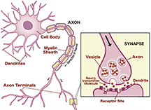
The dendrites of a neuron receive signals from other neurons. Upon receiving new information, the neuron transmits an electrical signal down the axon, triggering the release of chemicals called neurotransmitters that are deposited into a gap called the synaptic cleft between the terminals and the dendrites of the next neuron. The neurochemical crosses the synaptic space and activates receptors on the postsynaptic membrane to activate or inhibit the neuron’s electrical activity.
- Axons – extends from the cell body to transmit information
- Axon Hillock - a bridge between the axon and the cell body. The action potential, which begins at the axon hillock, is an electrical impulse that travels down the axon toward the terminal button
- Dendrites – specialized treelike or feathery extensions that branch from the neuron into the immediate area of the cell body
- Terminal Buttons – specialized structures which produce neurochemicals
- Synapse – the tiny gap between two neurons
- Pruning - a complex, regulated, activity dependent process in which connections that are non-essential to the formation of the developing brain are hewed
- Myelin Sheath - a wrapping of myelin around axons that serve as an electrical insulator that speeds nerve impulses
- Glial Cells - cells that provide support to neurons
- Oligodendrocytes – myelinate axons in the CNS
- Astrocytes – ‘star’ cells that support and guide neurons during development and act as ‘sinks’ for charged ions involved in neural activity, and remove neuroactive and potentially neurotoxic substances from the extracellular environment; form scars in the CNS
- Nodes of Ranvier - periodic gap in the insulating sheath (myelin) on the axon of neurons that serves to facilitate the rapid conduction of nerve impulses (see saltatory conduction)
- Resting Potential - the potential across the membrane when the cell is at rest (when there is no signaling activity). The resting potential for a neuron is approximately -70 mV. Electrical charge is unevenly distributed between the inside and outside of a neuron, with the inside being more negative under normal resting conditions.
- Action Potential - part of the process that occurs during the firing of a neuron. Part of the neural membrane opens to allow positively charged ions inside the cell and negatively charged ions out. This process causes a rapid increase in the positive charge of the nerve fiber. When the charge reaches +40 mv, the impulse is propagated down the axon.
- Synaptic Cleft – the space between the pre- and postsynaptic components (dendrite or spine of another neuron)
- Synaptic Vesicles – contain neurotransmitters or neuromodulators which are released by the presynaptic terminal into the synaptic cleft
- Saltatory Conduction – action potential ‘jumps’ from node to node between the myelin sheathes
- Neurotransmitters – permit the exchange of information among neurons. Each neurotransmitter has a specific shape – a ‘key’ that must fit a ‘lock’ on the receptor site. Specific neurotransmitters attach themselves only to a receptor with an appropriate fit. Neurotransmitters are classified according to their molecular size:
- Biogenic amines
- Acetylcholine (ACh)
- Serotonin (5-HT)
- Catecholamines
- Dopamine (DA)
- Norepinephrine (NE) (noradrenalin)
- Epinephrine (EPI) (adrenalin)
- Amino Acids
- Gamma-aminobutyric acid (GABA)
- Glycine
- Glutamate
- Aspartate
- Histamine
- Peptides
- Vasopressin
- Oxytocin
- Thyrotropin-releasing hormone (TRH)
- Corticotropin-releasing factor
- Substance P
- Tachykinins
- Cholecystokinin
Activating Systems:
Cholinergic System (Acetylcholine)
- Plays a role in normal waking behavior
- Associated with memory by maintaining neuron excitability
- Decrease in ACh in the neocortex has been associated with Alzheimer’s disease
Dopaminergic System (Dopamine)
- The nigrostriatial dopamine pathway takes part in coordinating movement and maintaining normal motor behavior
- The loss of dopamine neurons from the substantia nigra is associated with muscle rigidity and dyskinesia in Parkinson’s disease
- Dopamine release in the mesolimbic pathways causes feelings of reward and pleasure
- Excessive dopamine activity is associated with schizophrenia
- Affected by addictive drugs
Noradrenergic System (Norepinephrine)
- Norepinephrine is associated with learning by stimulating neurons to change their structure
- Facilitates normal development of the brain and plays a role in organizing movements
- Active in maintaining emotional tone
- Decrease in norepinephrine is associated with depression
- Increase in norepinephrine is associated with mania
Serotonergic System (Serotonin)
- Active in maintaining wakefulness
- Serotonin plays a role in learning
- Symptoms of depression is associated with decreases in the activity of serotonin neurons
- Increases of serotonin has been associated with schizophrenia and obsessive-compulsive disorder and tics
- Studies suggest an associated link between abnormalities in serotonergic nuclei and conditions such as sleep apnea and sudden infant death syndrome (SIDS)
Brain Structure and Function
 |
BrainVisor – Protect Your BrainBrainVisor does not provide medical or psychological advice, diagnosis or treatment recommendations. The material on this site is for informational purposes only and is not a substitute for your doctor or other health care professional's care.
|
 |
||
|


